Posts

Showing posts from November, 2016

Unilateral Maxillary Lateral Incisor Agenesis with Mini Implant Prostheses

Image
Many terms can be used to describe missing teeth. Anodontia is the complete absence of teeth; Oligodontia or partial anodontia means absence of six or more teeth; hypodontia denotes missing teeth, but usually less than six and often the size and shape of remaining teeth are altered as well, congenitally missing teeth or agenesis is defined as teeth that failed to develop or are not present at birth. Agenesis of any tooth can cause dental asymmetries, alignmentdifficulties, and arch length discrepancies but when the missing tooth is in the anterior region of the maxilla, the discrepancies can be quite noticeable. The maxillary lateral incisor is the second most frequently missing tooth after the mandibular second premolar even though Muller et al. found that maxillary lateral incisors experience the most agenesis (not including third molars). Agenesis of the maxillary lateral incisor is also linked with anomalies and syndromes such as agenesis of other permanent teeth, micro

The Hydrabrush/30 Second Smile Tooth Brush- Improving Gingival Health in Less Time, A Randomized Clinical Trial

Image
Plaque removal is the key to reducing the incidence of gingivitis, and the appropriate use of either a manual or an electric toothbrush is vital in achieving this goal. Electric toothbrushes have been shown in some cases to be more effective in removing plaque than manual brushes. The rationale behind the electric toothbrush is that it substitutes the hand coordination required with a manual toothbrush; with the electric toothbrush’s own cleansing mechanism. Electric toothbrushes can increase oral hygiene compliance for all ages, both for those with dexterity concerns and for children too . All that is required of the operator is turning on the toothbrush and then placing it at the desired brushing location. The major shortcoming of traditional electric toothbrushes is one of patient compliance. Considering each individual’s unique oral health condition and the time and the effort required for maintaining good oral hygiene, removing plaque via tooth brushing is a challenge .

Vitamin D Nuclear Receptor and Periodontal Disease

Image
Vitamin D has received increasing attention in recent years beyond its customary role in bone metabolism. There has been an explosion of scientific evidence which suggests the profound role that vitamin D plays in immune homeostasis. Vitamin D nuclear Receptor (VDR) has been identified as a ligand-dependent transcription factor which, in concert with its endogenous ligand, 1,25hydroxivitamin D (1,25(OH)2D3), is involved in the expression of countlessgenes.  In view of recent research which revealed that most immune cells express the vitamin D nuclear receptor (VDR), the authors reviewed the current knowledge regarding the immunomodulatory effects of vitamin D through its VDR on peridondontal disease. This manuscript provides a viable link between immune responses to periodontal pathogens and VDR and proposes a possible immunomodulatory treatment strategy for fine-tuning the host’s immune response against periodontopathogens that would diminish the destructive aspects of the r

Patient Satisfaction and Complications of Over-the-Counter Bleaching Products

Image
Hydrogen peroxide (HP), the active agent in bleaching products can, depending on the reaction conditions, form (per-)hydroxyl anions, superoxide anions and other radicals. Both HP and free radicals can diffuse various distances into tooth substance depending on their lifetimes and reaction environment. Although the action mechanism is not fully understood, the radical-involved oxidativeprocess can cause breaking of molecule bonds in stains in dentin and enamel. The resulting products appear less colored.  Bleaching products that are administered by the patient at home, either initially supervised by a dentist or many types that are purchased over-the-counter (OTC), contain carbamide peroxide (CP) to increase product stability. In an oral, watery environment, CP is decomposed to 36% HP and 65% urea. HP further decomposes into water and oxygen, while the urea breaks down to ammonia and carbon dioxide. Ammonia may contribute to a beneficial basic solution which can increase th

Healthy Lifestyle For Healthy Gums

Image
Due to improper practice of dental hygiene, bacteria in the mouth form plaque on teeth. Biofilm formed by colonizing bacteria that accumulate naturally on the teeth release toxic substances that harm the gums and cause them to become infected. Infection and inflammation that result when body attacks the bacteria can degrade gums and surrounding bone. One may experience exceptionally swollen and painful gums that are likely to bleed. Plaqueand associated microflora have been widely accepted as the primary cause of gum disease. However, another school of thought believes that the biofilm character of plaque allows for survival of a diverse flora, including less acid-tolerant organisms, some of which can produce ammonia from arginine or urea to counter acidification. Evidence is presented by certain authors that both supragingival and subgingival plaque have active oxygen metabolism and that plaque bacteria, including anaerobes, have developed defenses against oxidative stress.

Dental Ailments may be risky for Cardiac Patients

Image
Patients that are suffering from cardiac t=disorders are susceptible to physical and mental stress. Dental ailments add to the stress further as these patients are unable to cope with the pain due to cardiac disorders. These patients may encounter medical emergencies in case of dental disorders and they may demand multi disciplinary approach to achieve the success. Additionally, the results of cardiac surgery may suffer if the oral cavity is not maintained well since there is always a fear of infection affecting the heart from the oral cavity. Hence, the cardiac surgeon has to refer surgical patients to a dentist for oral prophylaxis and to treat any oral infections before cardiac surgery to prevent infective endocarditis. PDF LINK

Group Psycho Therapy prevents weight regain after Bariatic Surgery

Image
Success of the weight loss surgeries depends on certain factors like nutritional intake, psychological and surgery related factors may affect them so that they regain weight. Weight loss after the surgery may also brings with it several cognitive and behavioral challenges to these patients. Group psychotherapy may help in restoring the body image as well as the emotional balance of the patients. 30 patients: mean age=25-26; 18 women, 12 men; mean Body Mass Index (BMI) 48 SD ±8.31suffering from onset obesity, were enrolled in this study. They had five years of various and unsuccessful diets. They were assessed for bariatric surgery: sleeve or gastric by-pass. Patients were referred to the Eating Disorder and Obesity Unit of the School of Medicine “Federico II” Naples by the Bariatric Regional Units. They signed a written voluntary informed consent form before entering the study. All participants were evaluated before bariatric surgery in according to the standard procedure fo

Esthetic and Functional Rehabilitation of Molar Incisor Hypomineralisation: Report of Two Cases

Image
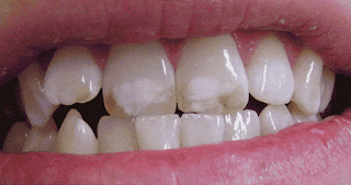
Molar-Incisor-Hypomineralisation (MIH) is a qualitative defect of 1-4 first permanent molars with or without the maxillary and mandibular permanent incisors. It seems to have been recognized first in the 1970s and prevalence varies between 2.8% and 25%, dependent upon the study. The term molar incisor hypomineralization (MIH) was introduced in 2001 to describe the clinical appearance of enamel hypomineralization of systemic origin affecting one or more permanent first molars (PFMs) that areassociated frequently with affected incisors.   Earlier condition was referred as hypomineralized PFMs, idiopathic enamel hypomineralization, dysmineralized PFMs, non-fluoride hypomineralization and cheese molars and is attributed to disrupted ameloblastic function during the transitional and maturational stages of amelogenesis. MIH’s clinical management is challenging due to: 1) the sensitivity and rapid development of dental caries in affected PFMs; 2) the limited cooperation of a youn

The Challenge of Treating Oral Infections Caused by Biofilms

Image
In natural systems, the harsh environments, nutrient/respiration needs, and sheer flow, make a community lifestyle preferable or inevitable for microorganisms. Furthermore, the bacteria take advantage of the communities′ organization to protect them from eradication by conventional antimicrobial therapy. A biofilm community is a microorganism aggregate characterized by cells that are attached to a substratum or interface or to each other; are embedded in a matrix of extracellular polymeric substances (EPS) that they have produced; and exhibitan altered phenotype with respect to growth rate and gene transcription.  The oral cavity is an example of a challenging environment for the long-term persistence of bacteria and fungi. Fluctuations in nutrient supply, temperature, pH, and the shear force of saliva flow have selected for a biofilm community adapted to high cell density, species diversity, and dynamic growth conditions. The microorganism production of EPS matrix is also ve

Knowledge and Public Perception of Dental Fluorosis in Children Living in Palestine

Image
Dental fluorosis is an enamel defect caused by excessive fluoride intake during the period of enamel formation, leading to incomplete crystal growth and increased porosity. Dental fluorosis is the first visible sign indicating that much fluoride has poisoned the whole body. Beside the health hazards there are other adverse effects of dental fluorosis such as high costs of other medical anddental problems and the effect on the victims and their loss of time. Rare knowledge of the causes of dental fluorosis may be considered one of associated risk factors which increase dental fluorosis problem because it will be reflected on people practice and attitude. Different studies have found very low knowledge on risk factors of dental fluorosis and its disadvantages. Regarding public perception, previous studies showed that more than half the children who have dental fluorosis were avoiding smiling and not accepting their appearance. In addition, parents felt worried and embarrassed wh

The Proper Diagnostic Pathway of Gardner’s Syndrome: Role of the Dentist in the Early Detection of the Associated Systemic Pathologies

Image
Gardner’s syndrome, a variant of Familial Adenomatous Polyposis (FAP), is a genetic disorder with highly penetrant autosomal-dominant transmission (80-100%). The incidence of FAP is approximately 1:7500 births, while Gardner’s syndrome occurs in 1:14000 births.  The first researcher reporting a clinical case characterized by the presence of manifestations which could be ascribed to Gardner’s syndrome was Fitzgerald in 1943, however, the combination of these signs was recognized as a genetically determined syndrome only in 1953 by Gardner e Richards.  This disease is characterized by the following triad: Gastrointestinal polyposis, with a 100 percent chance of malignant degeneration, other signs are bone tumors, frequently located on maxillary and cranial bones, and lastly the soft tissues tumors. PDF LINK

The Management of Crown Fracture of Immature Teeth by MTA and Calcium Hydroxide

Image
Traumatic injuries to teeth with or without pulpal involvement occur in children and adolescents. The majority of these injuries occur before root formation is complete, causing in some cases pulp inflammation and necrosis, with possible impact on the quality of life of affected individuals. Anterior crown fractures are favoured by the protruded position of maxillary incisors and their eruptive pattern. The most susceptible age to dental trauma is between 6 to 12 years . Complicated crown fractures represent 18-20% of all traumatic injuries topermanent teeth.  The treatment options vary due to tooth maturity, the time lapse between the accident and the treatment, the severity of pulp exposure, the presence or absence of haemorrhage, the size of the remaining crown, periodontal status and occlusal relations .  Root formation is classified in seven stages by Moorrees et al. Vital amputation (pulpotomy) is the treatment of choice for traumatized immature teeth with pulp exposure.

Unusual Root Staining of the Third Molars in a Patient Exposed to Lead and Tetracyclinec

Image
Intrinsic staining of teeth can result from introducing a variety of substances into the body which can form complexes with hard tissues (enamel, dentine, and bone). Two well-known substances that can cause such intrinsic staining of the teeth are lead and tetracyclineclass medications. Lead has been used for centuries in many industrial processes such as the development of batteries, sheets for X-Ray films, protective gear for high-radiation purposes (lead doors and lead gowns), and more stigmatically in the past, gasoline and paint. Lead poisoningoccurs when a person of any age swallows, absorbs, or inhales lead in any form.  Tetracycline and some of its homologues, such as minocycline, have a tendency to deposit in the hard substance of tooth and bone by forming a tetracycline-calcium-orthophosphate complex, which produces an intrinsic stain. The intensity of the stain depends on the dosage, duration of treatment, and age of onset of treatment. The age of onset of the tre

Keratocystic Odontogenic Tumor Involving Coronoid Process and Condyle

Image
Originally, KCOT was referred as Odontogenic keratocyst (OKC). It was first described by Hans Phillipsen in 1956 and its characteristic features were elaborated by Pindborg and Hansen in 1963. The term OKC was changed to ‘Keratocystic Odontogenic tumor’ by the World health organisation (WHO) in 2005. This change in nomenclature was mainly based on its potential for high recurrence, genetics and its neoplastic behaviour. KCOT is a benign uni or multicystic neoplasm with characteristic features of parakeratinised lining with stratified squamous epithelium.  This tumor which was once considered as an odontogenic cyst arises from the remnant cell rests of the dental lamina. In this article, a thorough literature review along with a case discussion is presented . A 25 year old female patient reported to the department of Oral medicine with the complaint of pain in the lower back tooth region since 3 months and a swelling on the same region (left lower 1/3rd of face) since 1 month. T

Enostosis Mimicking a Supernumerary Tooth

Image
A 17 year old female with history of malaligned teeth was referred from orthodontist for extraction of a supernumerary tooth along with first premolars in lower jaw. This is a usual practice to gain space for future orthodontic treatment. The referral included an orthopantomogram revealing a dense, well defined radiopaque lesion in relation to the periapical region of lower left first and second premolars. It is horizontal in alignment measuring 2 cm inlength and 5 mm in width lying parallel to lower border in close proximity withroots of premolars. On examination, clinically there was no cortical bone expansion or enlargement on the labial or lingual side.  The patient did not show any signs or symptoms of infection. The orthodontist considered this radiopaque lesion (supernumerary tooth in jaw) to interfere with his orthodontic treatment or potentially cause resorption of roots.  In view of determining the proximity of this radiopacity to surrounding structures, we undertook

Herbal Formulation (Tooth Paste): Anti-Precancerous Properties

Image
Toothpaste can be employed as a vehicle to improve and prevent oral health of individuals and population. Different modalities of treatment are offered for different dental and oral diseases, but the major drawback of these conventional drug therapies are side effects associated with their use. This has led to the interest in the discovery of novel anti-infective natural compounds.  The formulation is based on polyherbal extracts which display no side effects on the oral cavity as well as other body organs. To confirm the performance of the toothpaste formulation, the technology (Oral Sure, Orange Herbs Pvt Ltd, India) was evaluated in vivo on 20 patients (oral leukoplakia, oral lichen planus, submucous fibrosis and erythroplakia). Valid diagnostic tests were evaluated for diagnosing oral pre-cancerous lesions. Read more.............

Papillon-Lefevre Syndrome in 9 Year Old Pediatric Patient

Image
Papillon-Lefevre syndrome (PLS), 1st described by two French physicians Papillon and Lefebvre in 1924, is an extremely rare genodermatosis inherited as an autosomal recessive trait, affecting children between the ages 1-4 years. The disorder is characterized by diffuse palmoplantar keratoderma and premature loss of both deciduous and permanent teeth. The second major feature of PLS is severe periodontitis, which starts at age 3 or 4 years. The development and eruption of the deciduous teeth proceed normally, but their eruption is associated with gingival inflammation and subsequent rapid destruction of the periodontium. After exfoliation, the inflammation subsides and the gingiva appears healthy. Nail changes are apparent in advanced cases, manifested by transverse grooving and fissuring. The cause of PLS is not well understood, research groups have reported that loss-of-function mutations affecting both the alleles of the cathepsin-C gene, located on chromosome 11q14.1-q14.

A Case Report Using Oral Appliance Therapy Plus Oropharyngeal Exercise

Image
Snoring and obstructive sleep apneas are common disorders that affect at least 40 million Americans over 40 years of age. It is becoming more common in recent years for dentists to learn to fabricate and treat patients who have Obstructive Sleep Apnea with oral appliances. However, Oral Appliance Therapy often yields incomplete outcomes. Adjunctive therapeutic modalities may then be deployed in order to improve the overall effectiveness of therapy.The cause of sleep disordered breathing is a collapse of the pharynx that occurs with the onset of sleep and loss of muscle tonus in the upper airway.  It has been shown that pharyngeal musculature and function is altered in patients with OSA and that one of the contributing factors in the progression of OSA is thought to be progressive muscle weakness with age. Oropharyngeal exercise (OP) has been shown to improve moderate OSA in a group of Brazilian men using a complex set of exercises based on speech therapy practices. In this cl

Periodontal Splinting with Ribbon

Image
One of the critical manifestations of periodontal diseases is the sequel of mobility that results from such a clinical situation. Mobility, as extremely slowly developing phenomenon, leads to drastic consequences (especially toothmigration and occlusal trauma) that can be corrected. Depending on clinical conditions, tooth mobility can be treated by combination of several treatment modalities, such as periodontal and restorative therapeutics.  The periodontic therapy is directed toward the etiologic factors including plaque, and calculus. Root planning and subgingival debridement are performed to help to reduce inflammation, and bleeding. A few months after initial debridement, the tissue response is assessed. The periodontist will determine if the periodontal statue is stable enough to proceed with restorative treatment (splinting). Read more........................

Prevalence of Taurodont Primary Teeth in Turkish Children

Image
Taurodontism is a dental anomaly characterized by a significant shape abnormality that involves the roots. This anomaly is a condition that causes the pulp chamber or pulp tissue to enlarge and root bifurcation or trifurcation to migrate to the apices; this results from the shortening of the roots and root canals. The term taurodontism was first defined in 1913 by Sir Arthur Keith. It was subdivided into three types, as hypotaurodontism, mesotaurodontism and hypertaurodontism, depending on the relative amount of apical displacement of the floor of the pulp chamber. It appears both in permanent and deciduous teeth, but deciduous teeth are more frequently affected than permanent teeth. Get PDF Link

Influence of Repeat Pours of Addition Silicone Impressions on the Dimensional Accuracy of Casts

Image
Making an impression represents a critical step in processing and fitting of a dental prosthesis. The definite impression should be accurate to fabricate restoration with ideal marginal fit, internal fit, interproximal contacts and occlusal contacts. The impression materials are used to register or reproduce the form and relations of the teeth and the surrounding oral tissues. Dimensional accuracy and stability are theprimary requisites of an impression material.   Accuracy of an impression depends on properties of impression materials like thermal contraction, polymerization shrinkage, presence of volatile by products, elastic recovery, bulk of material and impression technique used. Other factors which influence the accuracy of an impression are tray material, space between tray and tooth preparation, storage conditions, relaxation of stresses caused by the use of non-rigid trays, excessive seating pressure, too slow removal from the mouth or an impression removed before th